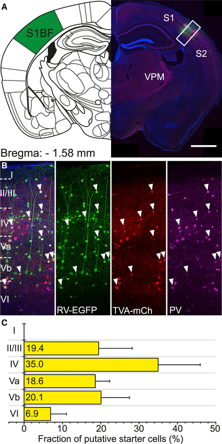
Figure 3. Identification of Starter Cells in Vgat-Cre/PV-Flp Mice.
(A) Coronal section through an injection site in right barrel cortex (scale bar, 1,000 μm).
(B) Inset in (A). Cells marked by white arrowheads are AAV-TVA-mCh and RV-EGFP co-transduced, putative starter cells. They are almost entirely positive for PV protein (scale bar, 100 μm).
(C) Distribution of putative starter cells across cortical layers (n = 12 mice; mean ± SD).

