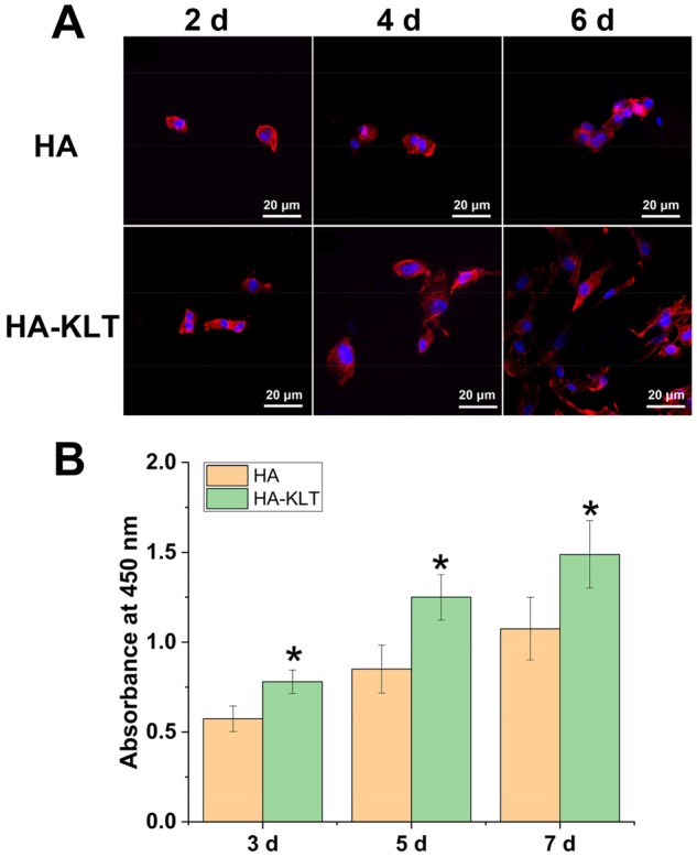
Figure 5.

Adhesion and proliferation of HUVECs on HA and HA-KLT gels. (A) LSCM image of HUVECs on the HA and HA-KLT hydrogel at 2, 4 and 6 days. Cells were stained with rhodamine phalloidin for actin and DAPI for nucleus. (B) Cell proliferation on the HA and HA-KLT gels by CCK8 assay after 3, 5 and 7 days. Data are expressed as the mean ± standard deviation for n = 3. *P < 0.05
