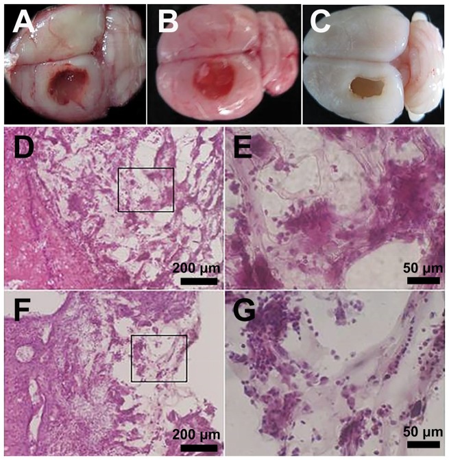
Figure 6.
In the left frontal cortex region of rats, lesion cavities (5 × 3 × 3 mm3) were mechanically created. The HA or HA-KLT gel was injected immediately to fill up the lesion cavities. The harvested brain tissue of HA group (A), HA-KLT group (B) and blank control group (C) after implantation for 4 weeks. Representative H&E staining of tissues embedded with HA gel were shown in (D) 10× and (E) 40× (indicated in black box) views. H&E staining of tissues embedded with HA-KLT gel were shown in (F) 10× and (G) 40× (indicated in black box) views

