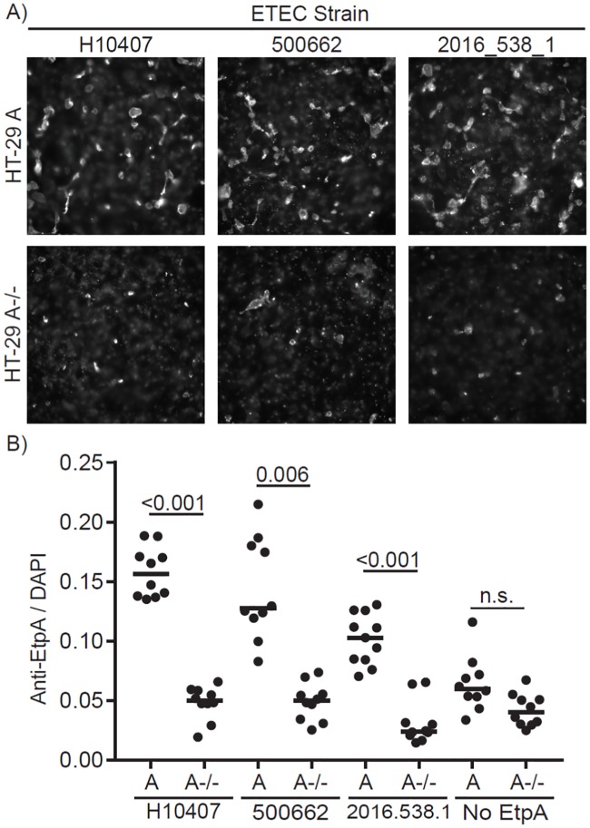
Fig 4. EtpA function is conserved.
(A) Immunofluorescence microscopy of EtpA bound to HT-29 WT cells expressing Blood Group A sugars or HT29 A-/- CRISPR deletion mutant generating functional blood group O cells, EtpA detected with anti-EtpA antibodies. (B) Quantitation of mean fluorescent values normalized to DAPI (nuclei) signal using Volocity software. Statistical differences determined by Kruskal-Wallis testing followed by Dunn’s test for multiple comparisons with p<0.05 considered significant.

