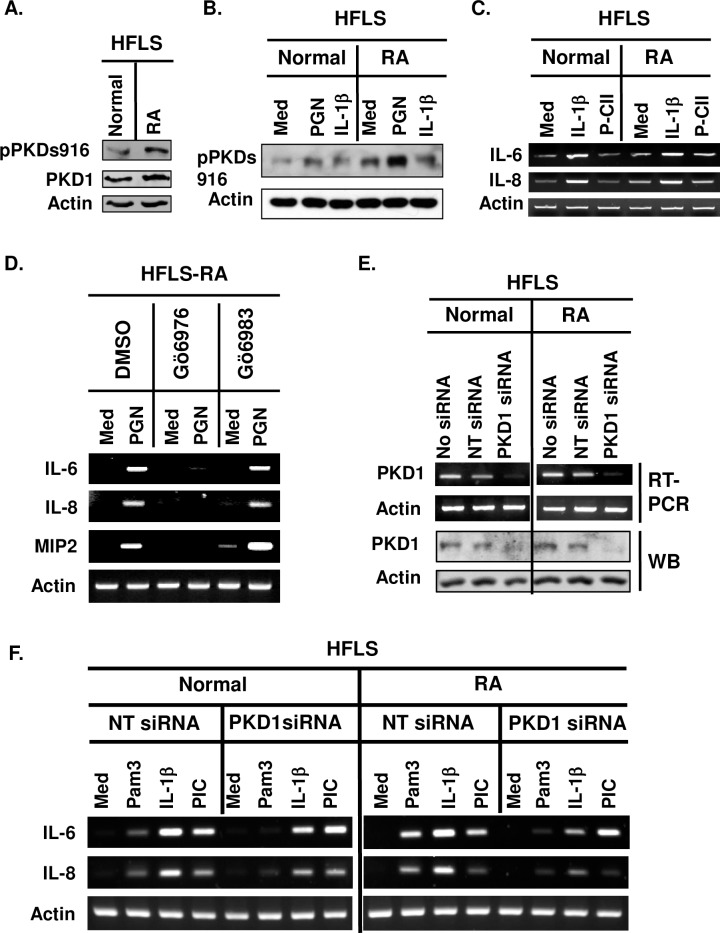
Fig 1. PKD1 can be activated and plays an essential role in both spontaneous and TLR/IL-1R-induced expression of proinflammatory mediators in human synoviocytes.
(A) Protein levels and phosphorylation status of PKD in human primary fibroblast-like synoviocytes (HFLS) from a normal donor (HFLS-N) and from a patient with RA (HFLS-RA) were detected by Western blot. (B) HFLS-N and HFLS-RA were stimulated with media (med), TLR2 ligand peptidoglycan (PGN; 50 μg/ml), or human recombinant IL-1β (10 ng/ml) for 45 min. Protein levels and phosphorylation status of PKD were detected by Western blot. (C) HFLS-N and HFLS-RA were stimulated with media (med), IL-1β (10 ng/ml), or synthetic CII peptide (P-CII; 50 μg/ml) for 4 hr. Levels of IL-6 and IL-8 mRNA were analyzed by RT-PCR. (D) HFLS-RA were pretreated with vehicle (1% v/v DMSO), Gö6976 (500 ng/ml) or Gö6983 (500 ng/ml) for 1hr and then stimulated with PGN (10 μg/ml) for 4 hr. Levels of the indicated cytokine and chemokine mRNA were analyzed by RT-PCR. (E, F) HFLS-N and HFLS-RA were transiently transfected with non-target siRNA (NT siRNA; control) or PKD1-specific siRNA (PKD1 siRNA; PKD1-knockdown). mRNA levels (E-top panel) and protein levels (E-bottom panel) of PKD1 were detected by RT-PCR and Western blot, respectively. Control and PKD1-knockdown HFLS were stimulated with Pam3CSK4 (Pam3; 500 ng/ml), IL-1β (10 ng/ml), or TLR3 ligand PIC (50 μg/ml) for 4 hr. Messenger RNA levels of the indicated gene were analyzed by RT-PCR. All experiments were repeated two to three times with similar results. S1 Fig shows uncropped blot and gel scans.

