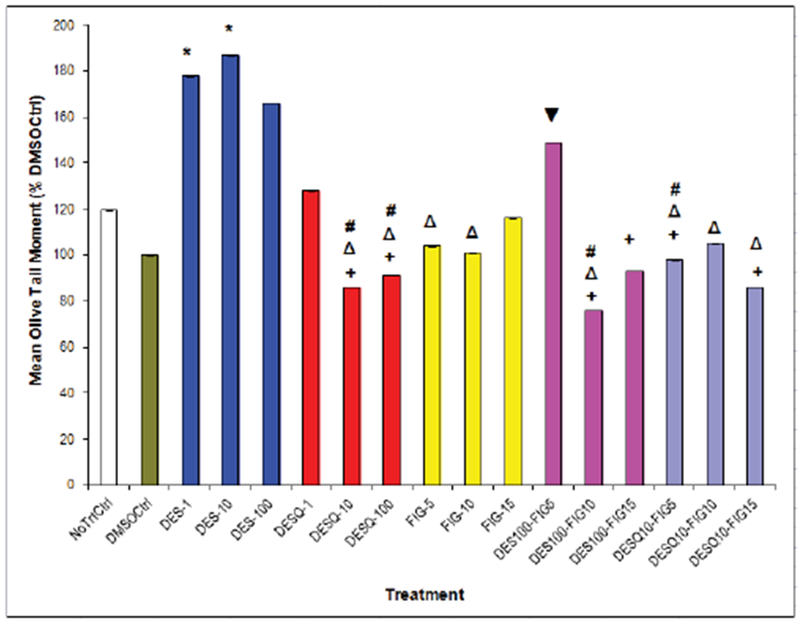
Figure 5:

Chemically-induced DNA Damage in Benign Human Breast Epithelial (MCF10A) Cells as Measured by Comet Assay.
Effects of DES, fig extract, or combined treatments on MCF10A cells. Induction or attenuation of DNA damage in human breast epithelial (MCF10A) cells with DES (0.1 – 10 μM), fig extract (5 – 15 μL), or high-dose DES plus fig combinations for up to 6 h.
NoTrtCtrl = No treatment control; DMSOCtrl = Dimethyl sulfoxide preserved control; DES = Diethylstilbestrol; FIG = Ficus carica leaf extract; DES-1, DES-10, DES-100 = DES 1, 10, and 100 μM, respectively; FIG-5, FIG-10, FIG-15 = FIG 5, 10, and 15 μL, respectively.
* Compared to DMSO control, p<.05
+ Compared to DES-10, p<.05
Δ Compared to DES-1, p<.05
# Compared to DES-100, p<.05
▼ Compared to DES 100-Fig 10
