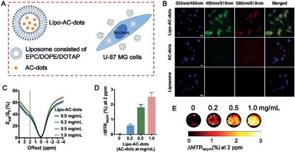
Figure 3.
Liposome encapsulation of AC-dots and cellular uptake by human U-87 MG glioma cells. A) Schematic illustration of the cellular uptake of AC-dots following liposome encapsulation. B) Fluorescence imaging of cells after 6 h incubation with Lipo–AC-dots, AC-dots, or empty liposome. Nuclei are counterstained with DAPI. Scale bar=20 μm. C–E) CEST MRI measurements of cells labeled with Lipo–AC-dots at different concentrations (37°C, B0=11.7 T). Data in D: mean±SD (n=3).

