
Fig. 2. Carboxylated PBAE nanoparticles mediate cytosolic protein delivery.
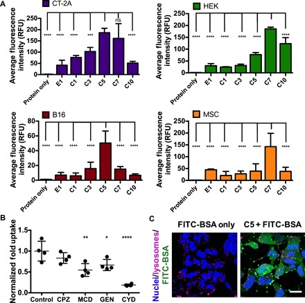
(A) Average fluorescence intensity of cells treated with carboxylated PBAE nanoparticles encapsulating FITC-BSA (300 ng of FITC-BSA per well, 20 w/w). Data are presented as means + SD (n = 4); statistical significance is determined by one-way ANOVA with Dunnett’s post hoc tests comparing uptake levels to that of the nanoparticle formulation achieving the highest levels of FITC-BSA uptake in each cell line. ***P < 0.001 and ****P < 0.0001. (B) Uptake by HEK cells in the presence of different endocytosis inhibitors. CPZ, chlorpromazine; MCD, methyl-β-cyclodextrin; GEN, genistein; CYD, cytochalasin D. Data are presented as means ± SD; statistical significance is determined by one-way ANOVA with Dunnett’s post hoc tests as compared to the control group (n = 4). *P < 0.05, **P < 0.01, and ****P < 0.0001. (C) Confocal images of HEK cells treated with C5/FITC-BSA nanoparticles or protein alone for 4 hours. Scale bar, 10 μm.