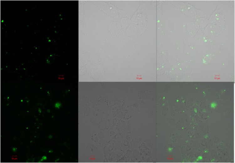
FIG. 8.
Confocal microscopy of HUVECs incubated on a μ-Slide I Luer perfused with tumor media (ES-2 top panels, U87 bottom panels) with CFSE-labeled MVs. The left panels correspond to the fluorescent detection channel, middle panels are brightfield detection channel, and the right panels are the combined images.

