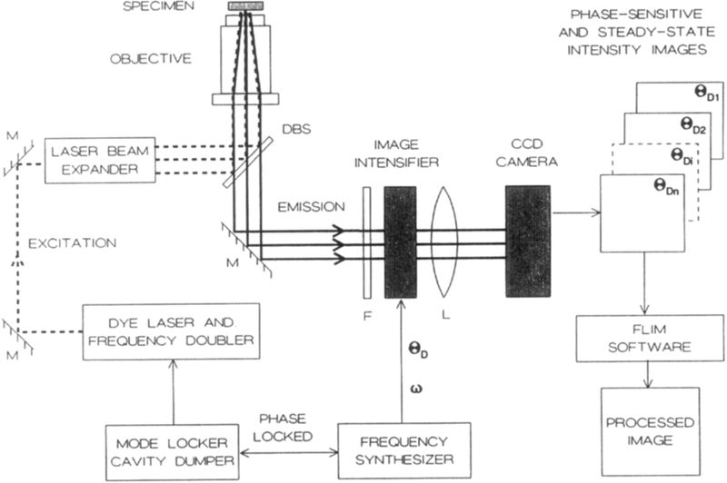
Fig. 1.
Instrumentation for fluorescence lifetime imaging microscopy. The excitation is presently the frequency-doubled output of a pyridine-1 dye laser, which is synchronously pumped by a mode-locked Nd:YAG (neodymium:yttrium-aluminum-garnet) laser and cavity dumped at 3.81 MHz. The excitation light is expanded by a Newport LC075 (10×) laser beam expander. A Nikon Diaphot-TMD inverted fluorescence microscope is equipped with a Nikon Fluor 40×, NA 1.3 objective and DM400 Nikon dichroic beam splitter (DBS). The gated image intensifier (Varo 510-5772-310) is positioned between the target and the CCD camera. The gain of the image intensifier is modulated using the output of a PTS 300 synthesizer with digital phase shift option. The detector is a CCD camera (Photometrics, series 200, thermoelectrically cooled PM 512 CCD).

