Figure 3.
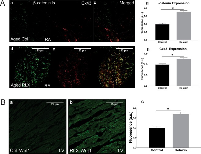
Age and relaxin’s effect on the levels of Cx-43 and β-catenin in aged atria. (Aa–c) Aged right atrial tissue (Aa–c) expressed significantly less β-catenin and Cx43 than RLX-treated animals (Ad–f). (Ag–h) Quantification of atrial β-catenin and Cx43 expression (n = 5/group). 60x magnification. (B) RLX (Bb) increased Wnt1 expression in LV compared to control (Ba). (Bc) Quantification of Wnt1 expression in LV sections. 600x magnification, scale bars = 25 µm and apply to all panels.

