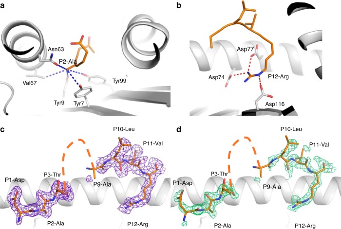
Fig. 1.
Structure of the HLA-A*68:01-NP145 complex. Structure of the HLA-A*68:01 (white cartoon) bound to the NP145 peptide (orange stick). a A zoomed view of the P2-Ala anchor residue interaction with the HLA-A*68:01 molecule, the blue dashed lines represent hydrophobic interactions. b The P12-Arg salt bridge network (red dashed lines) with the HLA-A*68:01 amino acids. c, d The electron density maps after refinement (c 2Fo-Fc map colored in purple and contoured at 1 σ) or before building the peptide in d (Fo-Fc or omit map colored in green and contoured at 3 σ). The orange dashed line on c, d represent the missing amino acid from the NP145 peptide (P4–P8) in the crystal structure.

