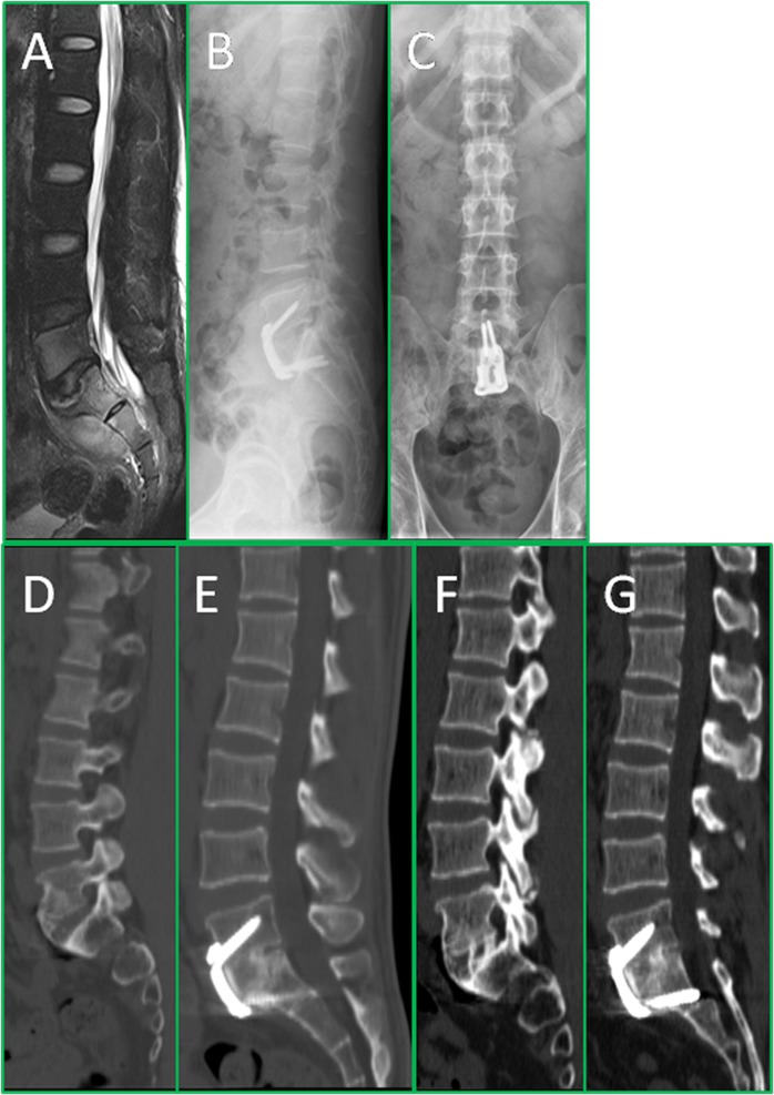
Figure 3.
The series of radiographs show the radiological changes before and after treatment. This is a 35-year-old male patient, who had L5-S1 spinal TB with pre-sacral abscess, low back pain, and right lower limb radicular pain. The pre-operation MRI (A) shows bone destruction of L5 and S1 and pre-sacral cold abscess formation; The post-operative X-ray (B,C) confirmed the position of plate; The 12 months post-operative X-ray (D,E) shows definite bone fusion, while intervertebral height and the lumbosacral angle maintained satisfactory correction; The 24 months post-operative X-ray (F,G) reveals the ARCH plate still in position and good inter-body fusion of L5-S1, and the intervertebral height and lumbosacral angle were well maintained.

