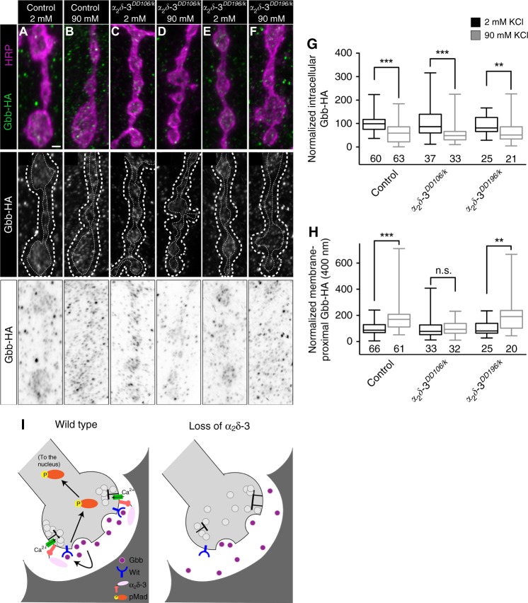
Fig. 10. α2δ-3 limits diffusion of Gbb following its activity-dependent release.
a–f Top: representative z-projections of boutons of the indicated genotypes labeled with HRP (magenta) and Gbb-HA (green). Genotypes specifically are: control (D42>gbb-HA), α2δ-3DD106/k (α2δ-3DD106/k10814; D42>gbb-HA), and α2δ-3DD196/k (α2δ-3DD196/k10814; D42>gbb-HA). Scale bar: 1 µm. Middle: individual Gbb-HA channel shown in grayscale. Membrane-proximal Gbb-HA was measured between the two dashed white lines. Bottom: individual Gbb-HA channel with inverted colors. g Quantification of intracellular Gbb-HA normalized to control levels before (2 mM KCl) and after (90 mM KCl) neuronal stimulation. For all Gbb-HA release experiments, n is the number of NMJs scored. h Quantification of membrane-proximal Gbb-HA (within 400 nm) normalized to control levels before and after neuronal stimulation. n.s. not significantly different. **p < 0.01; ***p < 0.001 by a two-tailed Mann–Whitney test. i Proposed model for α2δ-3 acting as a physical barrier to promote Gbb signaling. Upon release from the neuron, Gbb is released into the synaptic cleft. With the aid of α2δ-3, Gbb remains in close proximity to the presynaptic membrane and is then able to activate the BMP Type II receptor Wit. The receptor complex in turn phosphorylates the transcription factor Mad, transducing a BMP signal back to the nucleus of the neuron.

