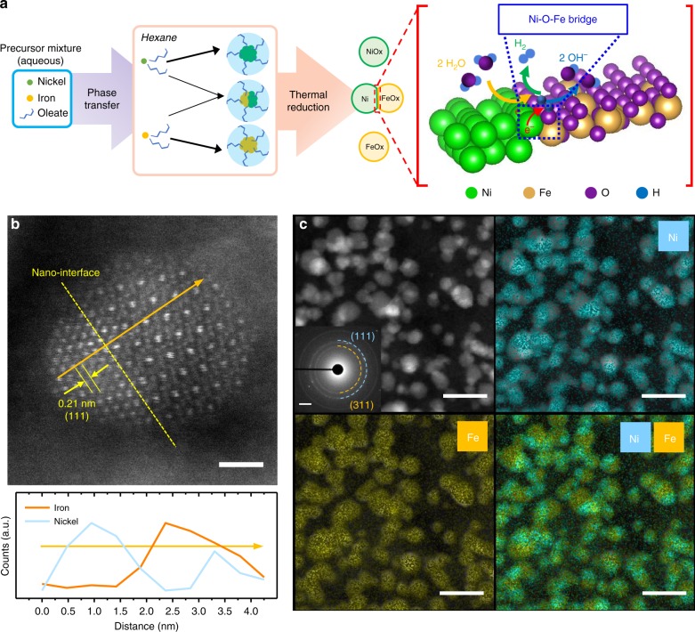
Fig. 1.
Nanoparticle design and electron microscopies. a Schematic representation of the Ni and Fe nanoparticles and the Ni-Fe Janus nanoparticles synthesis through the oleate-assisted micelle formation and the illustration on the HER across the Ni-γ-Fe2O3 interface in alkaline medium. b STEM-HAADF image of a single Ni–Fe NP nanoparticle and its corresponding EDS line-scan spectrum (scale bar: 1 nm). c High-resolution EDS mapping on STEM-HAADF images of the nanoparticles for Ni and Fe, selected area electron diffraction inset (image scale bars: 20 nm; SAED scale bar: 2 nm−1).

