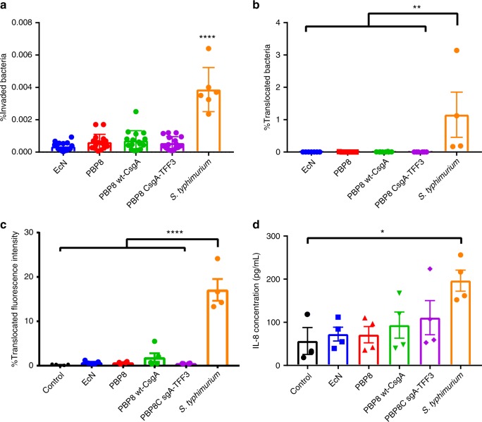
Fig. 3. Effects of curli fiber expression on EcN pathogenicity.
a Percent of bacteria that invaded a monolayer of Caco-2 after 2 h of co-incubation with EcN, PBP8 variants, and S. typhimurium. b Bacterial translocation to the basolateral compartment of polarized Caco-2 cells exposed to bacterial library for 5 h. c Epithelial permeability of polarized Caco-2 24 h post-infection, quantified via FITC-dextran (MW 3000–5000) translocation. d IL-8 secretion from the basolateral compartment of polarized Caco-2 cells 24 h post-infection. Data are represented as mean ± SEM. (ns) p > 0.05, *p ≤ 0.05, **p ≤ 0.01, ***p ≤ 0.001, ****p ≤ 0.0001, one-way ANOVA followed by Tukeyʼs test. Source data are provided as a Source Data file.

