Figure 1.

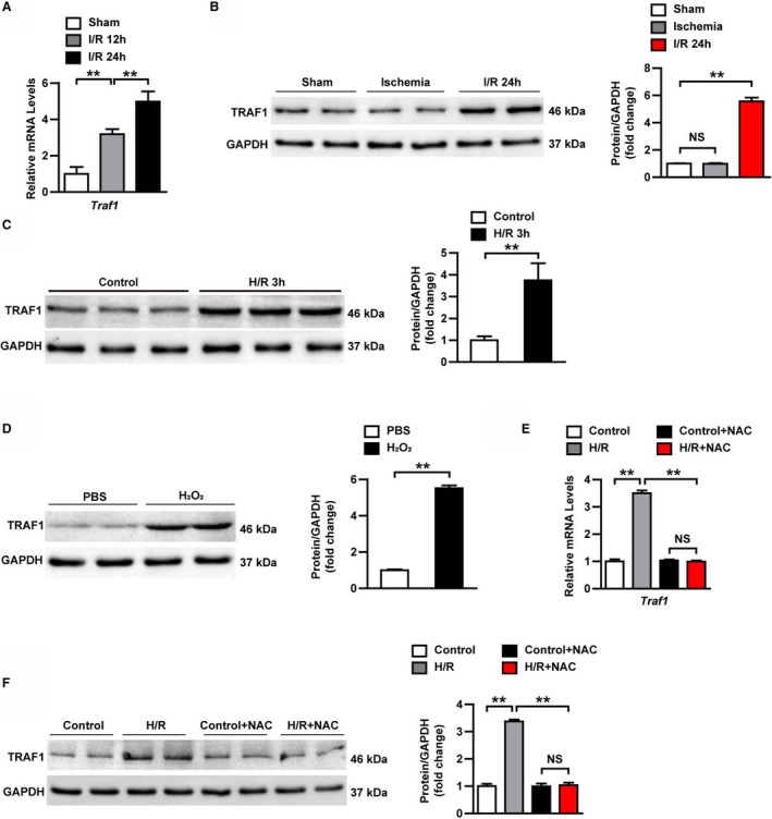
TRAF1 expression is increased by reactive oxygen species during myocardial I/R. A, mRNA expression level of Traf 1 in heart samples of mice at indicated points after I/R (n=3 per group). B, TRAF1 protein expression level in heart tissues in the indicated groups (n=4 per group). C, TRAF1 protein expression level in neonatal rat primary cardiomyocytes exposed to H/R. D, TRAF1 protein expression level in neonatal rat primary cardiomyocytes exposed to H2O2. E, mRNA level of Traf 1 in neonatal rat primary cardiomyocytes in the indicated groups. F, TRAF1 protein expression level in neonatal rat primary cardiomyocytes in the indicated groups. Results shown are representative of 3 blots. For panels (B, C, D, and F), GAPDH served as loading control. For statistical analysis, 1‐way ANOVA was used for panels (A, B, D, and E); a 2‐tailed Student t test was used for panels (C and D). **P<0.01. H/R indicates hypoxia/reoxygenation; I/R, ischemia/reperfusion; NAC, N‐acetyl‐L‐cysteine; NS, not significant; TRAF1, tumor necrosis factor receptor–associated factor 1.
