Figure 1.

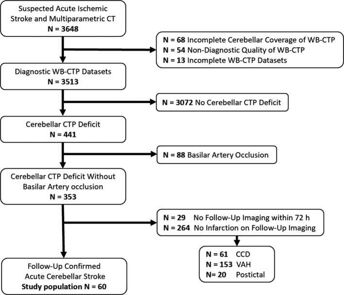
Flow chart of patient selection. CCD indicates crossed cerebellar diaschisis; CT, computed tomography; VAH, vertebral artery hypoplasia; WB‐CTP, whole‐brain CT perfusion.

Flow chart of patient selection. CCD indicates crossed cerebellar diaschisis; CT, computed tomography; VAH, vertebral artery hypoplasia; WB‐CTP, whole‐brain CT perfusion.