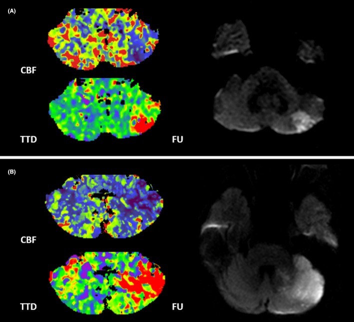
Figure 2.

Case examples. Patient examples of acute cerebellar stroke. Patient A is a 77‐year‐old man with an initial CBF deficit volume of 5 mL of the left cerebellar hemisphere. On follow‐up MRI, the patient had a PICA territory infarction with a FIV of 2 mL. Patient B is a 67‐year‐old woman with an initial CBF deficit of 21 mL of the left cerebellar hemisphere. On follow‐up MRI, the patient had a PICA and SCA territory infarction with a total FIV of 22 mL. Both patients received IVT. CBF indicates cerebellar blood flow; FIV, final infarction volume; FU, follow‐up MRI; IVT, intravenous thrombolysis; PICA, posterior inferior cerebellar artery; SCA, superior cerebellar artery; TTD, time to drain.
