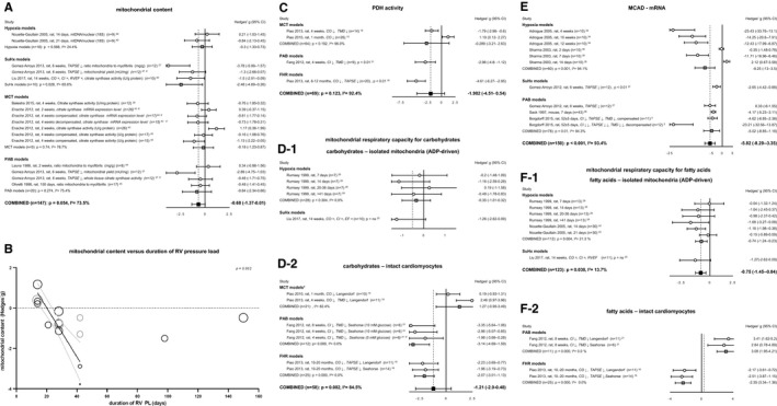
Figure 5.

Mitochondrial function. Plots of mitochondrial content measured by mentioned methods (A). Bubble plot showing relation between mitochondrial content and duration of RV PL (B). Forrest plot of PDH activity as reflection of mitochondrial breakdown of pyruvate to acetyl‐CoA (C). Forrest plots of mitochondrial respiratory capacity for carbohydrate metabolites measured in isolated mitochondria (ADP‐driven) (D‐1) or intact cardiomyocytes (D‐2). Forrest plots of MCAD expression at mRNA level (E), as representative of the β‐oxidation. Forrest plots of mitochondrial respiratory capacity for fatty acids measured in isolated mitochondria (F‐1) and intact cardiomyocytes (F‐2). Data are presented as Hedges’ g. Combined Hedges’ g are presented as squares: gray representing Hedges’ g of a specific model, black representing Hedges’ g of all included studies. Bars represent 95% CI. Bubble size represents relative study precision, calculation based on SD. Gray bubbles are not included in meta‐analysis. Black line represents regression line, gray lines represent 95% CI. = indicates not statistically significant affected. ↓, decreased; ↓↓, decreased compared with decompensated group; CI, cardiac index; CO, cardiac output; FHR, fawn hooded rats; I2, level of heterogeneity; MCT, monocrotaline; n, number of included animals; PAB, pulmonary artery banding; PDH, pyruvate dehydrogenase; PL, pressure load; RVEF, RV ejection fraction; TAPSE, tricuspid annular plane systolic movement; X, not included in meta‐analysis. *Significantly (P<0.05) increased compared with PAB.
