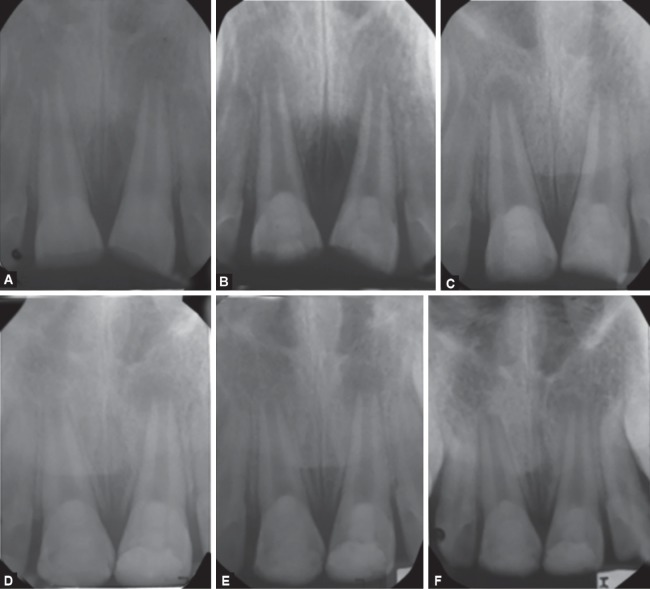
Figs 2A to F.
Group I BC vs group II PRP: immature bilateral, fractured teeth (#11 and 21) with an open apex and large periradicular radiolucency in a 9-year-old boy. 11 was treated with PRP and 21 was treated with blood clot. Periapical radiographs: (A) Preoperative periapical radiolucent lesion with an open apex; (B) After the placement of mineral trioxide aggregate; (C) At the 3-month follow-up, partial regression of periapical radiolucent lesion; (D) At the 6-month follow-up, with marked reduction in periapical lesion with continued development of the root; (E) At the 9-month follow-up, nearly complete healing of periapical lesion with continued development of the root apex; (F) At 1-year follow-up, complete maturation of the root apex

