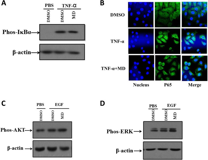
Fig. 5.
The activations of NF-κB and MAPK signaling pathways and ICP0 promotor activity were not affected by MD. a HeLa were pretreated with DMSO or MD (3.0 μM) for 1 h prior to stimulation with TNF-α (100 ng/mL) for 15 min. Then some of the HeLa cells were subjected to western blot to analyze the occurrence of IκBα phosphorylation. The rest of HeLa cells were stained with anti-P65 antibody (green) and DAPI (blue). The pictures were captured by confocal laser scanning microscope. b RD cells were cultured in serum free medium for 5 h. Then cells were treated with DMSO or MD (3.0 μM) and cultured in serum free medium for additional 1 h. EGF was added for 15 min to stimulate activation of MAPK pathway. The occurrence of AKT (c) and ERK (d) phosphorylation were analyzed by western blot analysis

