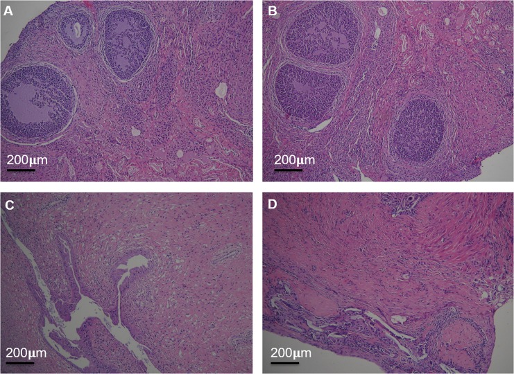
Figure 7.
The reproductive organs were examined by HE staining under light microscopy. The structure of the ovary of saline group (A) and (PEI-SA)HA/PC group (B); the structure of the uterus of saline group (C) and (PEI-SA)HA/PC group (D). There is no significant difference in ultra-structure between the control group and the (PEI-SA)HA/PC group.

