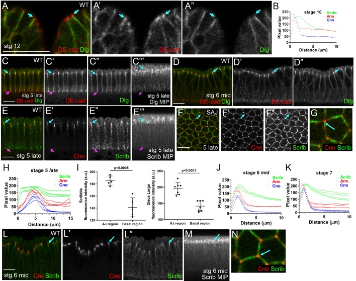
Fig. 1.
Scrib and Dlg localize to nascent AJs during cellularization. (A-A″) Stage 12 ectoderm. Dlg localizes to septate junctions, basal to AJs. (B) Quantification at stage 10. Pixel plots along the apical-basal axis reveal separation. (C-J,L-N) Cellularization to early gastrulation. Dlg (C-D″) and Scrib (E-G,L) localization relative to SAJs. (C-E″,L) Cross-sections. (F-G,N) En face sections through SAJs at the level of highest enrichment. (C‴,E‴) Maximum intensity projections (MIPs). (C-C″,E-E″) Dlg and Scrib localize along the basolateral membrane during cellularization, overlapping both SAJs (cyan arrows) and BJs (magenta arrows). (F) At the SAJ level, Scrib is uniformly distributed around the cell circumference without TCJ enrichment (G,N, cyan arrows). (D-D″,L-L″,M) Gastrulation. Dlg and Scrib remain enriched near apical SAJs (cyan arrows). (H,J,K) Quantification via pixel plots reveals the changing localization of Scrib relative to AJ proteins. (I) Quantification of relative levels of cortical Scrib and Dlg at the SAJ level versus basolateral. Data are mean±s.d. with individual data points indicated. Scale bars: 10 µm.

