Fig. 2.

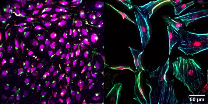
Primary pulmonary artery endothelial cells. Representative images of nuclei (magenta), actin (blue), eNOS (green) and VEGF (red) in (left) normal PAEC and (right) PPHN PAEC.

Primary pulmonary artery endothelial cells. Representative images of nuclei (magenta), actin (blue), eNOS (green) and VEGF (red) in (left) normal PAEC and (right) PPHN PAEC.