
Fig. 7.
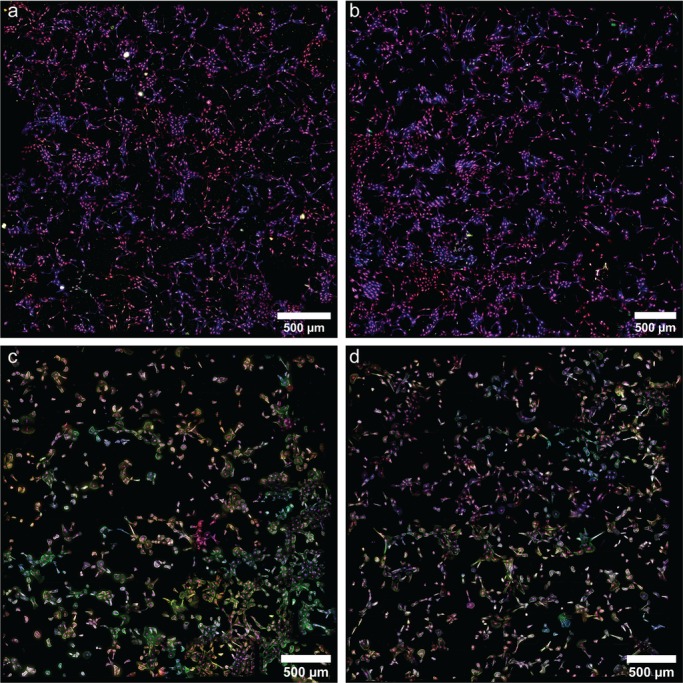
Tube formation assays in normal and PPHN PAEC. Representative high-throughput imaging assays of nuclei (purple), actin (blue), eNOS (green) and VEGF (red) in 18 h tube-formation assays for (A) normal PAEC in vehicle media, (B) normal PAEC in IGF-1 media, (C) PPHN PAEC in vehicle media and (D) PPHN PAEC in IGF-1 media. Two replicates performed from different cell isolations for each normal and PPHN PAEC are presented here.