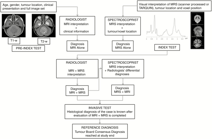
Fig. 1.
Study Protocol and Workflow for Readers of 1H-Magnetic Resonance Spectroscopy. 1H-magnetic resonance spectroscopy (MRS) was interpreted independently by 1) radiologists and 2) an expert spectroscopist. All readers were blinded to the reference standard of the tumor board consensus diagnosis and final histopathology. Radiologists determined diagnosis in 2 stages: MRI interpretation in combination with clinical information, and MRS interpretation in combination with MRI results and clinical information (index test). The spectroscopist performed a similar process: MRS interpretation blinded to clinical and radiological information (but knowing tumor location and voxel position), and MRS interpretation with the differential diagnosis made by radiologists.

