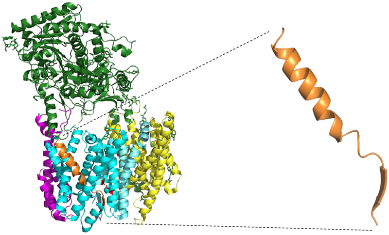
Figure 6. Structure of an APP substrate bound to the γ-secretase complex.

Newly reported cryo-EM structure of the γ-secretase complex bound to an APP substrate (Rendered from PDB ID: 6IYC). One of the active site aspartates is mutated to alanine, and both substrate and presenilin were mutated with cysteine to allow disulfide crosslinking. Blue: Presenilin; Yellow: Aph-1; Magenta: Pen-2; Green: Nicastrin; Orange: APP substrate. APP substrate alone is also shown to illustrate that the conformation of bound substrate resembles that of designed TMD substrates.
