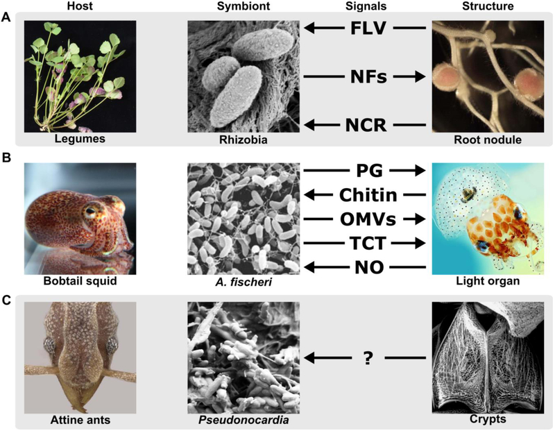
Figure 1. Signal-structure interfaces in bipartite associations between (A) legumes and rhizobia, (B) Hawaiian bobtail squid and A. fischeri, and (C) attine ants and Pseudonocardia.
The arrows indicate the direction of signals from producer to respondent and are arranged from top to bottom in each panel to indicate the order of signaling. FLV, flavonoids; NF, nodulation factors; NCR, nodule-specific cysteine-rich peptides; PG, peptidoglycan; OMVs; outer membrane vesicles; TCT, tracheal cytotoxin; NO, nitric oxide. Image credits: legume, rhizobia, and root nodule, Jean Michel Ané; bobtail squid, Mark Mandell; A. fischeri from [28]; light organ from Spencer Nyholm, under the terms of the Creative Commons Attribution License; attine ant, Ted Schultz; Pseudonocardia, Cameron Currie; ant crypt, Cameron Currie.

