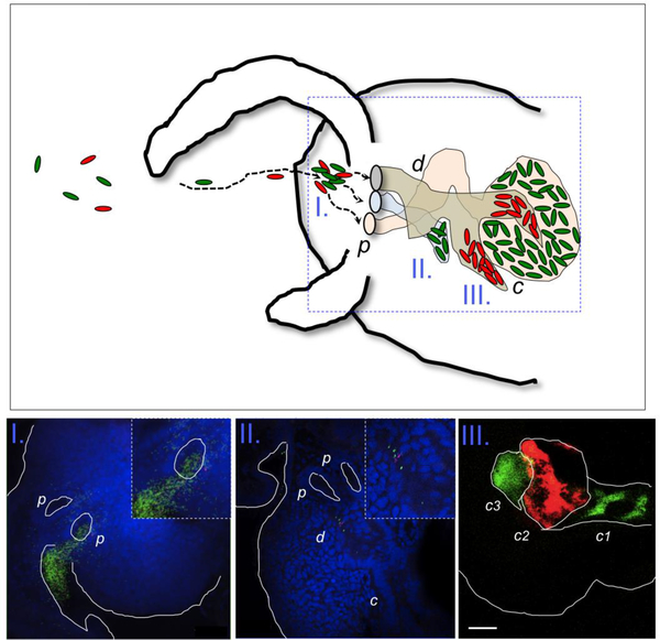
Fig. 1.
(Upper) Different V. fischeri strains, present in the environment, (I.) form aggregates around the three pores (p) of the light organ, then detach and (II.) migrate through the duct (d), and finally (III.) arrive and colonize deep in the crypts (c) of the light organ. Green and red ovals represent different V. fischeri strains in the environment, and the dashed arrows are their trajectories. (Lower) Confocal images corresponding to the different stages of bacterial behavior along the colonization path. Bacteria are GFP or RFP labeled; nuclei of host tissue (blue) were stained with TOTO-3. Cells of V. fischeri are aggregated (in I.), migrating through the ducts (in II.), and colonizing the light organ crypts (in III.); bar = 50 mm. Note that each crypt is typically colonized by a single bacterium (green or red) that initiates the symbiont population therein.

