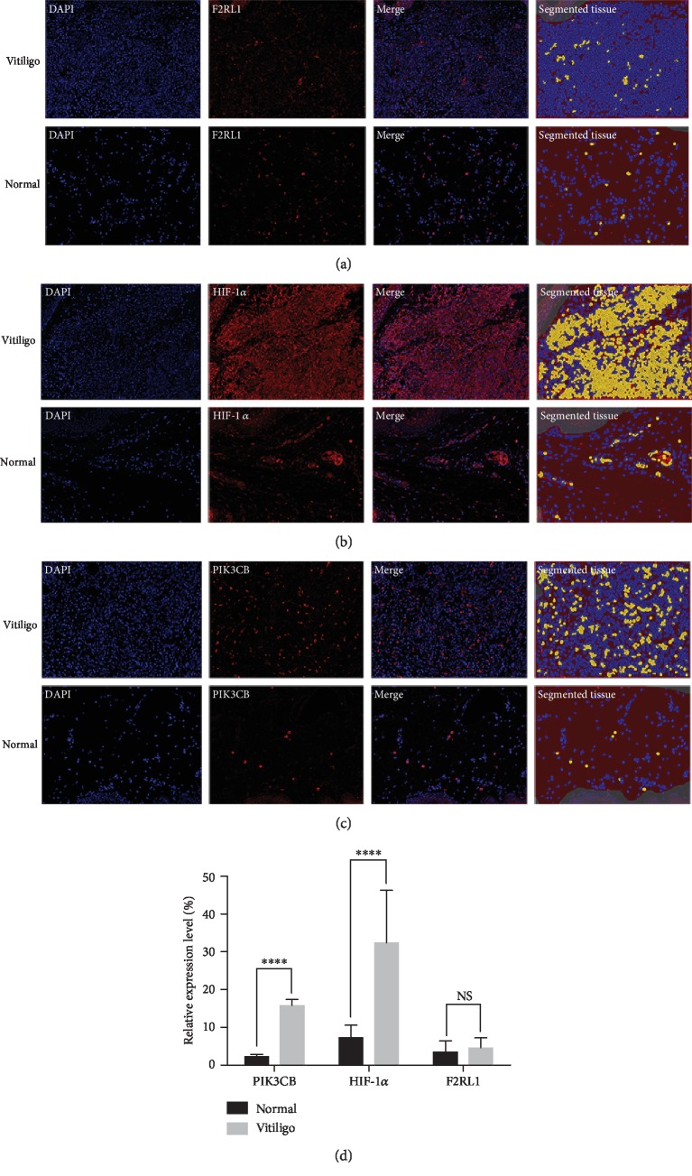
Figure 4.
Opal IHC validation of DEGs. F2RL1 (a) were nonsignificant in lesional skin of patients with vitiligo compared with normal control (n = 10). HIF-1α (b) and PIK3CB (c) were significantly elevated in vitiligo (n = 10). Three or more areas were randomly selected. Pictures were taken at ×200 and analyzed using inFORM image analysis software, which quantifies the segmented tissue based on respective positive expression (yellow). (d) Data are shown as mean ± SEM. ∗∗∗∗p < 0.0001. NS = nonsignificant.

