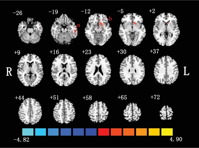
Figure 1.
Two-sample t-test was performed between non-NPSLE patients and healthy controls (p < 0.05, corrected). Warm colors exhibited increased mALFF in non-NPSLE patients compared to healthy controls, and blue colors exhibited the opposite (a: left inferior temporal gyrus; b: left putamen; R: right hemisphere; L: left hemisphere). This figure was created in Slice Viewer [67].

