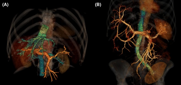
Figure 2.

3D‐reconstruction of intravenous contrast, the portal venous system is coloured orange and the caval venous system is coloured cyan. (Panel A) healthy control. (Panel B) patient with polycystic liver disease and hepatic venous outflow obstruction. No hepatic veins are visible due to external compression by cystic liver tissue. Renal transplant is also visible
