
Figure 3.
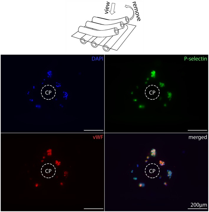
Crossing points (CPs) from low vWF‐loaded gas exchange fibers. The center of the CPs is empty but decorated with small aggregates consisting of a few nucleated cells (DAPI, blue), with P‐selectin‐positive (green) platelets in their cytoplasm (PLAs, platelet–leukocyte aggregates) as well as cytoplasmic and extracellular vWF‐spots (red) [Color figure can be viewed at https://wileyonlinelibrary.com]