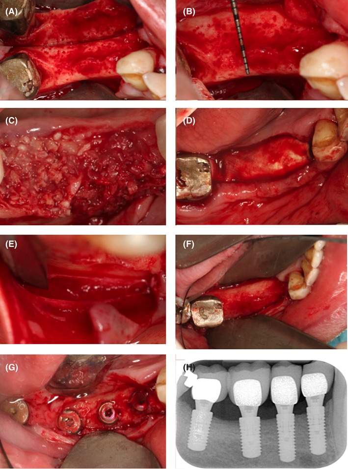
Figure 1.

Overview of the surgical procedure. A very thin alveolar crest appeared after reflection of a mucoperiosteal flap (A). The alveolar ridge was then prepared with cortical perforations (B). Thereafter, the bone allograft material soaked in the second phase of the PRGF solution was applied in order to build up the alveolar bone volume necessary for future implant placement (C). It was then covered with a resorbable collagen membrane (D). The surgical site was primarily closed by means of a periosteal incision (E), the use of horizontal mattress sutures and a continuous half‐hitch suture. The second surgical procedure took place after a healing period of 4 months. The significant gain in alveolar ridge volume can be appreciated (F). Four implants were placed according to the manufacturer's recommendations (Straumann Group, Basel Switzerland). Four months later, the implants were uncovered (G). Postoperative two‐dimensional radiographs demonstrate stable integration of the implants 36 months after final restauration (H)
