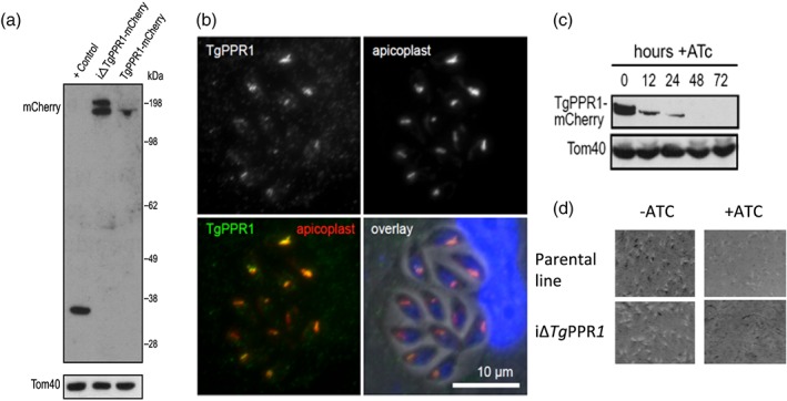
Figure 2.

Apicoplast protein TgPPR1 is necessary for parasite growth. (a) Western blot detection of mCherry‐tagged endogenous TgPPR1 using either the t7s4 promoter (iΔTgPPR1‐mCherry) or the native promoter (TgPPR1‐mCherry). The positive control is a Toxoplasma gondii cell line expressing mCherry. Tom40 acts as a loading control. The presence of two bands in the iΔTgPPR1‐mCherry lane is consistent with a preprocessed PPR targeting intermediate still bearing the predicted apicoplast targeting peptide and mature PPR‐mCherry fusion protein. The position of the 198 kDa standard is shown. (b) Colocation of TgPPR1‐mCherry expressed from the t7s4 promoter with resident apicoplast biotinylated proteins visualised by streptavidin staining. DNA staining in blue, TgPPR1‐mCherry in green, and streptavidin‐stained apicoplast in red. (c) ATc‐induced knock‐down of TgPPR1 assayed over 72 hr. Tom40 acts as a loading control. (d) Eight‐day plaque assay shows normal plaque formation in iΔTgPPR1 cells without ATc‐induced TgPPR1 depletion, but no plaques with ATc treatment, indicating that PPR is essential for normal growth. A control of the parental cell line is also shown
