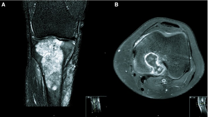
Figure 1.

A, Magnetic resonance imaging of case 1. A coronal T2 SPIR sequence of the right knee shows a multiloculated lesion in the proximal tibia epimetaphysis. The lesion contains T2 hyperintense cartilage nodules, which are typical for chondroid tumours. The extension into the epiphysis (in this case up to the subchondral bone plate) is not typical for a chondroid tumour, which is usually contained in the metaphysis. Small internal foci of low signal intensity are noted, and are in keeping with chondroid matrix calcifications. Perilesional bone marrow oedema and soft tissue oedema are present. B, Magnetic resonance imaging of case 2. Axial T1 SPAIR after administration of gadolinium contrast in the left distal femur shows a typical septonodular or ‘rings‐and‐arcs’ enhancement in this cartilaginous tumour. The lesion extends into the epiphysis and through the posterior cortex into the soft tissues. Perilesional bone marrow oedema is present.
