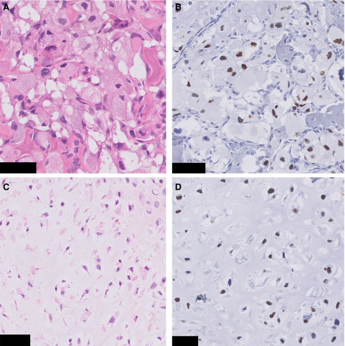
Figure 5.

p53 immunohistochemistry (case 3). A, Clear cell chondrosarcoma component. B, Immunohistochemistry for p53 in the same tumour region shows overexpression in the tumour cells. C, Conventional chondrosarcoma area. D, Immunohistochemistry for p53 shows overexpression in the tumour cells. Scale Bar: 50 μm (A–D).
