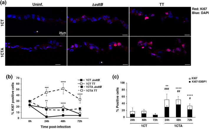
Figure 8.

Infection with genotoxic Salmonella promotes exit from quiescence of the 1CTA cells. 1CT and 1CTA cells, grown in 3D culture, were infected with the MC1 TT or MC1 ∆cdtB strain. Induction of proliferation status and DNA damage was assessed by immunofluorescence analysis, using antibodies specific for Ki67 and 53BP1, respectively, at the indicated time points. (a) Representative scanning confocal micrograph of infected 1CT and 1CTA cells grown in 3D culture stained with the anti‐Ki67 specific antibody (red). Nuclei were counterstained with DAPI (blue). Magnification 40×. The white asterisk indicates nuclei of fibroblasts embedded in the collagen matrix. (b) Quantification of cells positive for Ki67. (c) Quantification of cells positive for Ki67 (white bar) and double positive for both Ki67 and 53BP1 foci (black bar). Mean ± SEM of three to four independent experiments. Statistical analysis was performed using the Student t test. *,# p < .05; **,## p < .01; ***,### p < .001; ****,#### p < .0001. *comparison Ki67 positive cells, #comparison Ki67‐γH2AX double‐positive cells
