Figure 3.

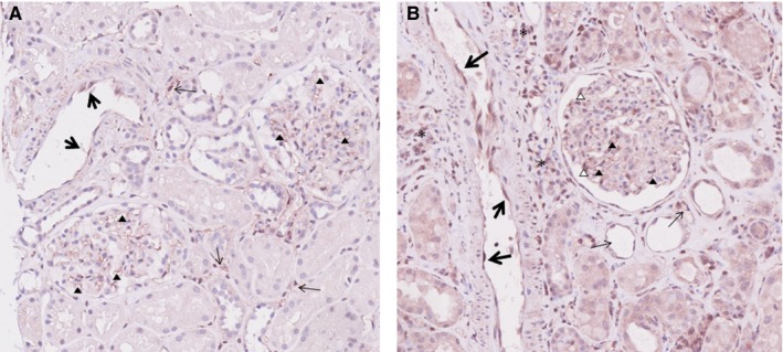
ARHGDIB expression in the kidney. A, In a transplanted kidney without histological abnormalities, weak ARHGDIB expression is seen in endothelial cells of interlobular arteries (large arrows), endothelial cells of peritubular capillaries (small arrows), and endothelial cells of glomerular capillaries (arrow heads). B, In a transplanted kidney with acute tubular necrosis, strong ARHGDIB expression is seen in endothelial cells of interlobular arteries (large arrows), endothelial cells of peritubular capillaries (small arrows), and endothelial cells of glomerular capillaries (solid arrow heads). In addition, positive staining for ARHGDIB is also seen in some podocytes (open arrow heads) and lymphocytes (asterisks) [Color figure can be viewed at http://www.wileyonlinelibrary.com]
