
Figure 2.
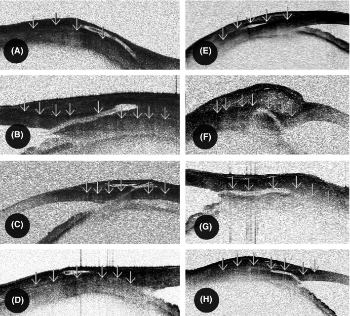
Position of the outer XEN Glaucoma Gel Microstent (XEN‐GGM) lumen in the conjunctiva visualized by OCT. As shown by Howlett et al., Tenon's layer (‘↓’) in the OCT appears as a hyperreflective section as opposed to the hyporeflective section underlying the sclera. The conjunctival stroma in OCT consists of irregular fibres, perfused blood vessels and cystic spaces and therefore appears more irregular. A–D were classified to the intraconjunctival group (Tenon's layer below the outer stent lumen in OCT) and E–H to intra‐ and subtenon group (Tenon's layer above the outer stent lumen in OCT).