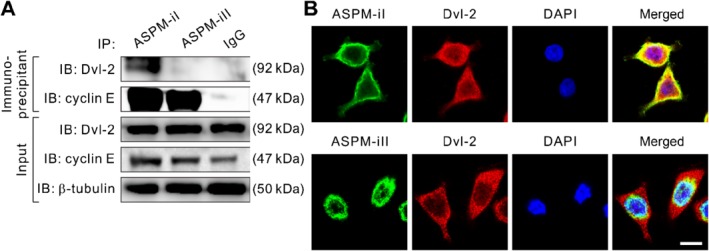
Figure 3.

ASPM‐iI and ASPM‐iII differentially interact with Dvl‐2 and cyclin E. (A) Co‐IP of ASPM‐iI or ASPM‐iII with Dvl‐2 or cyclin E using ASPM‐isoform‐specific antibodies or control IgG in NCKUH‐SP‐1 cells. β‐Tubulin was included as a loading control. (B) Representative confocal images showing the strong co‐localization (yellow) of ASPM‐iI (green) with Dvl‐2 (red) in NCKUH‐SP‐1 cells. Nuclei were counterstained with DAPI (blue). Note that ASPM‐iII does not co‐localize with Dvl‐2 (bottom). Scale bar = 10 μm.
