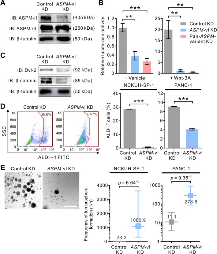
Figure 4.

ASPM‐iI specifically regulates Wnt activity and the stemness properties of PDAC cells. (A) IB showing the effect of the isoform‐specific KD of ASPM‐vI on the protein abundance levels of ASPM‐iI and ASPM‐iII in NCKUH‐SP‐1 cells. (B) Relative Wnt‐specific luciferase expression in control KD or ASPM‐vI KD and Wnt‐3a (250 ng/ml × 16 h)‐treated NCKUH‐SP‐1 cells. A non‐target shRNA (control shRNA) and an shRNA targeting all ASPM variants (clone TRCN0000118905; pan‐ASPM‐variant KD) were used as controls. (C) IB showing that specific KD of the expression of ASPM‐vI reduced the protein abundance levels of Dvl‐2 and β‐catenin in NCKUH‐SP‐1 cells. (D) ASPM‐vI KD diminished the population of ALDH+ cells in NCKUH‐SP‐1 cells. Representative flow cytometry plots showing the pattern of ALDH activity in control KD or ASPM‐vI KD NCKUH‐SP‐1 cells, with the frequency of the boxed ALDH+ cell population as a percentage of cancer cells shown. Bottom: the percentage of ALDH+ cells. (E) Representative phase contrast images of control KD or ASPM‐vI KD NCKUH‐SP‐1 cells. Scale bar = 200 μm. Right: limiting dilution assay demonstrating the tumorsphere‐forming efficacy of control KD or ASPM‐vI KD NCKUH‐SP‐1 cells. Mean ± SEM (n = 6 in each group). **p < 0.01; ***p < 0.001.
