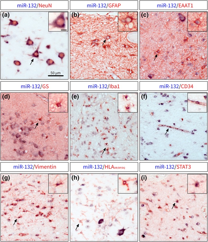
Figure 3.

Double labeling of miR‐132 with cell type‐specific markers in human TLE‐HS. (a)—miR‐132 was expressed in neurons throughout hippocampus, including granule cells, pyramidal cells and hilar neurons; co‐localization of miR‐132 was also found with markers of astrocytes GFAP (b), EAAT1 (c) and GS (d); miR‐132 was also co‐localized with Iba1+ microglial cells (e) in the areas of glial scar in CA1 and with CD34 (f) associated with blood vessels; the co‐localization with reactivity markers showed miR‐132 expression in vimentin+ cells with astrocytic morphology (g), HLA‐DR/DP/DQ‐expressing cells with microglial morphology (h) and STAT3+ cells in the CA1; black arrows indicate cells shown in higher magnification in insets; scale bar 50 μm; scale bar in inset a =10 μm and applies for insets a‐i [Color figure can be viewed at https://wileyonlinelibrary.com]
