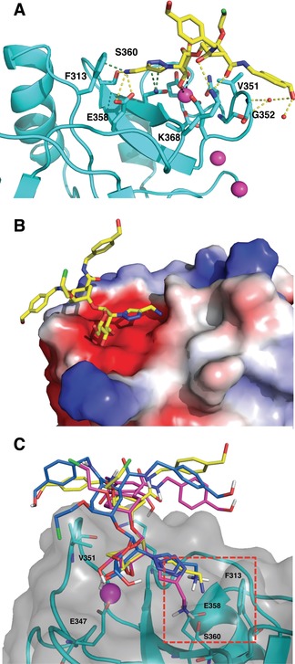
Figure 7.

A) Binding mode of 16 in the canonical Ca2+ binding site of DC‐SIGN (PDB 6GHV). H bonds are represented in yellow, van der Waals and cation–π interactions with Phe313 in green. Ca2+ ions are represented as magenta sphere. B) The same complex. View rotated by 180° and protein represented as electrostatic surface. C) 3D Structure of DC‐SIGN (cyan) in complex with compound 16 (blue) from MD simulation. Superimposed crystallographic (yellow, PDB 6GHV) and docked (magenta) poses of compound 16 are depicted.
