Figure 1.

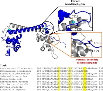
Structure of CueR (E.coli) (PDB id.: 1Q05‐modified) showing the potential metal binding sites (top). Sequence alignment of CueR proteins from various organisms (bottom). Conserved cysteine residues are highlighted in yellow.

Structure of CueR (E.coli) (PDB id.: 1Q05‐modified) showing the potential metal binding sites (top). Sequence alignment of CueR proteins from various organisms (bottom). Conserved cysteine residues are highlighted in yellow.