
Figure 5.
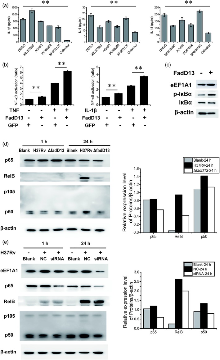
Mtb FadD13 and eEF1A1 regulate proinflammatory cytokines production by stimulating NF‐κB signalling pathway in macrophages. (a) ELISA results showing NF‐κB inhibitor (celastrol) inhibited the production of proinflammatory cytokines (IL‐1β, IL‐18 and IL‐6). Differentiated THP‐1 macrophages were infected at a MOI of 10 with M. smegmatis‐FadD13 for 6 hr. DMSO: DMSO treated macrophages, as the negative control; SB203580: p38 MAPK inhibitor; AG490: Janus kinase inhibitor; PD98059: ERK1/2 inhibitor; SP600125: JNK inhibitor; celastrol: NF‐κB inhibitor. The results presented as the one replicate from three independent experiments. * p < .05 and ** p < .01 (two‐tailed unpaired t test). (b) The results of dual luciferase reporter gene assay showing FadD13 activated NF‐κB signalling pathway in HEK293T cells. HEK 293T cells were transfected with pEGFP‐N1‐FadD13 or pEGFP‐N1 for 24 hr, respectively, then treated with TNF‐α or IL‐1β for 6 hr for activation the NF‐κB pathway. The results presented as the one replicate from two independent experiments. * p < .05 and ** p < .01 (two‐tailed unpaired t test). (c) Western blot results showing FadD13 promoted the phosphorylation of IκBα, the inhibitor of NF‐κB, in HEK293T cells. HEK 293T cells were transfected with pEGFP‐N1‐FadD13 or pEGFP‐N1 for 24 hr, respectively, followed by protein extraction and western blot. The results presented as the one replicate from two independent experiments. (d) Western blot results showing ΔfadD13 mutant decreased the expression of p65, p50 and RelB in the macrophages. Blank: uninfected THP‐1macrophages; H37Rv: H37Rv infected THP‐1 macrophages; ΔfadD13: ΔfadD13 infected macrophages. Macrophages were infected at a MOI of 10 with ΔfadD13 or H37Rv, respectively, for 1 and 24 hr. The right columns stand for the 24 hr western blot results. The results presented as the one replicate from two independent experiments. (e) Western blot results showing eEF1A1 decreased the expression of p65 and p50 (except RelB) in macrophages. Blank: normal THP‐1 macrophages; NC: THP‐1 macrophages transfected with control siRNA; siRNA: THP‐1 macrophages transfected with eEF1A1 specific siRNA. Macrophages were infected at a MOI of 10 with H37Rv for 1 and 24 hr. The right columns stand for the 24 hr western blot results. The results presented as the one replicate from two independent experiments