Figure 5.

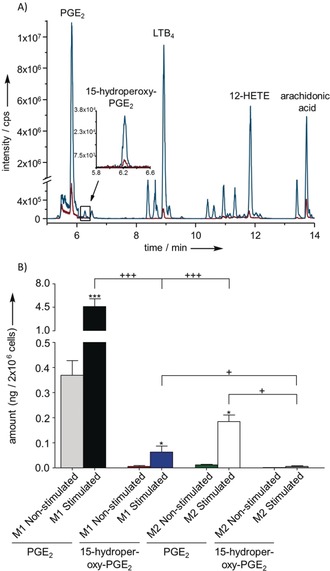
A) UHPLC‐MS profile of a lipid mediator extract from 2×10⁶ stimulated (blue) or non‐stimulated (red) human M1 macrophages measured in negative ionization mode, plotted as total ion count in full MS. The enhanced region shows the extracted ion chromatogram for 3. The novel PG 3 elutes at 6.25 min. Further known lipid mediators were detected: 1 at 5.82 min, leukotriene B4 (LTB4) at 8.93 min, 12‐HETE at 11.85 min, AA at 13.73 min. B) Amounts of 1 (grey, black, green, white) and 3 (red, blue, orange, dark grey) produced in 2×10⁶ stimulated (n=4) or non‐stimulated (n=3) human M1 or M2 macrophages shown as means ± SEM. Cells were suspended in 1 mL PBS plus 1 mm CaCl2 and incubated for 10 min at 37 °C with 2.5 μm A23187 or vehicle (0.5 % methanol). Statistical evaluation: one way ANOVA with Tukey Post‐hoc test, */+ P≤0.05; **/++ P≤0.01; ***/+++ P≤0.001, asterisks refer to comparison to non‐stimulated conditions.
