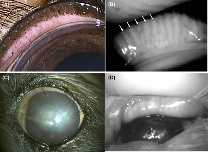
Figure 2.

Eyelid and meibographic findings for a dog of control group. Slitlamp findings of the externally rotated eyelid. (A) MG are not apparent. Meibographic findings using desktop meibography. (B) MG can be clearly observed (arrows). Corneal and meibographic findings for a dog of KCS group Extensive corneal vascularization (C), as well as meibographic findings (D) using portable type meibography, shortening findings (arrow head) are apparent
