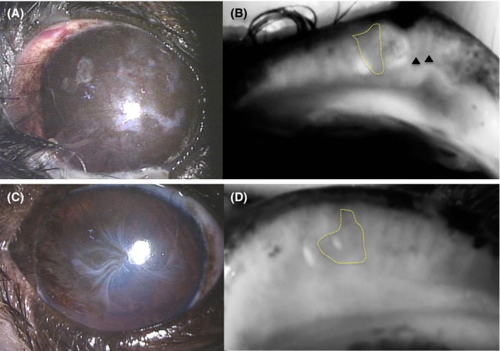
Figure 3.

Corneal and meibographic findings for two animals of the KCS group. Extensive corneal pigmentation and vascularization, (A) as well as meibographic findings using desktop‐type meibography of gland dropout (area surrounded by dotted line) and shortening (arrowheads) (B) are apparent. Corneal vascularization (C) and the meibographic finding using desktop‐type meibography of gland dropout (area surrounded by the solid line) in the center of the eyelid (D) are apparent
